

|
ReGena-Vet Laboratories, LLC |
|
The Best Medicine for Our Best Friends |
|
Tomorrow’s Cures Today |


|
What are MSCs? |
|
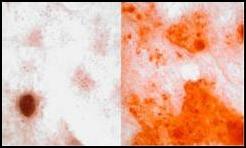
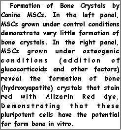
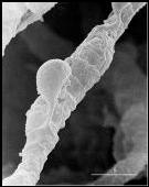
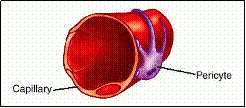
The unique properties of MSCs were first discovered by Alexander Friedenstein in 1976 (1). He reported that glass-adherent cells from bone marrow could synthesize bone crystals when grown under specified conditions. (figure to right). Since that initial discovery, much more has been discovered about these important cells (2-6). MSCs can differentiate into tendon, bone, cartilage, heart muscle, skeletal muscle, smooth muscle and other cell types in vitro (2-8). Although these cells are plastic (have the ability to express many proteins of other cell types), at RGVL we believe the major role of MSCs is to function as reparative cells and repair damaged tissues in vivo . Recent reports (7) have compared MSCs with pericytes that are found adjacent to capillaries (see the diagram and electron micrograph of a pericyte bound to a capillary on the lower right). Many believe that MSCs and pericytes are the same cell (8). Pericytes have been demonstrated to participate in the repair of injured endothelium, as have MSCs. A substantial case can be made for the identical nature of these two cells populations (8). However, the exact identify of MSCs will be a mystery until specific markers for MSCs are identified. |
|
Although a lot is known about the behavior of these cells in vitro (grown in tissue culture flasks), much less is known about their behavior in vivo (in living animals). MSCs have been tested and found to be effective in many animal disease models ranging from autoimmune disorders to myocardial infarction. However, the behavior of these cells in patients with real diseases is largely unknown. At ReGena-Vet Laboratories, our primary mission is to investigate the therapeutic potential of these medically important cells. |