


|
ReGena-Vet Laboratories, LLC |
|
The Best Medicine for Our Best Friends |
|
Tomorrow’s Cures Today |


|
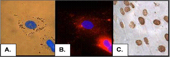
Techniques available for tracking injected MSCs. In panel A, the nuclei of canine MSCs have been stained with Hoechst 3352 DNA dye and the cytosol labeled with iron particles that allow tracing the cells using Magnetic Resonance Imaging. In panel B, the other MSCs have been stained with a red fluorescent dye that labels cytosolic granules. In panel C, MSCs pretreated with the DNA label, bromodeoxyuridine or BrdU have been stained with a monoclonal antibody against this marker. All of these techniques are available to locate MSCs that have been injected into animals. |
|
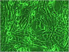
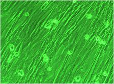
Photomicrograph of adult bone marrow stem cells (MSCs) from dog (above) and cat (below) magnified 200 fold. These cells were isolated from a patient’s bone marrow and the stem cells amplified over a million-fold by growth in special media. MSCs have been shown to improve many laboratory animal models of diseases. We believe that they will be equally effective in the canine and feline diseases as described below. |
|
To accomplish our mission, RGVL has developed many proprietary methods for producing therapeutic (large) quantities of these cells. In this past decade, ReGena-Vet has probably grown more dog and cat cells than any other laboratory in this field. In addition to large quantities of cells that we prepare regularly, our current and future studies require that we have the ability to trace the injected cells in vivo. This requires special labeling of the cells. To the right and below are photos that document our ability to grow and trace cells for our ongoing clinical research projects. |
|
Imaging MSCs: |