

|
ReGena-Vet Laboratories, LLC |
|
The Best Medicine for Our Best Friends |
|
Tomorrow’s Cures Today |


|
cells (9). Treatment with MSCs decreases fibrosis, increases cardiac output, cardiac function and survival. Similar result were obtained in laboratory rats using an autoimmune model of DCM (10). Taken together, these animal models demonstrate that MSCs will improve rodent DCM and suggest that they also have the potential to Dobes with DCM. |
|
In the example below, this Doberman’s fractional shortening (FS), as determined by echocardiography, was decreasing rapidly from above 20% to less than 13% in the two months just prior to stem cell treatment. Fractional shortening is the most common indicator used by veterinarians to indicate how hard the heart contracts and is determined during a “cardio” check at most dog shows. If this rate of decline had continued, it is estimated that euthanasia would have been necessary around |
|
Ongoing Clinical Trials 2 |
|
ReGena-Vet Laboratories, LLC has extensively modified these approaches to treat large dogs with DCM. First, a method of growing therapeutic (large) quantities of canine bone marrow stem cells from donors was developed. Then, a safe method for administering the MSCs into debilitated dogs with compromised circulatory system was developed. As a result of this research, several Dobes with DCM have been treated and all have shown improvement. |
|
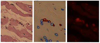
The heart from the first DCM Dobe treated with MSCs showed pronounced signs of DCM. Note the extensive white scar tissue in the left panel; the pink is cardiomyocytes (above left panel). Because the dog was so compromised (shortening fraction of less than 10%), the injected MSCs were labeled with a red fluorescent dye visualized in the unstained adjacent section (center and right panel). By merging the right panel onto the center panel, it is possible to demonstrate a close interaction of the cardiomyoctyes and injected red fluorescent MSCs. Clearly, the injected cells have migrated to the dog’s heart and appear to be interacting with the cardiomyocytes in areas of severe DCM pathology. We believe this interaction is beneficial, but more experiments are needed to verify this statement. Most importantly, these photomicrographs demonstrate that MSCs do “home” to areas of severe myocardial fibrosis and scarring. |
|
heritable form of dilated cardiomyopathy, are benefitted by treatment with adult bone marrow stem |